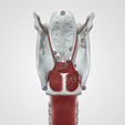
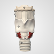
The larynx is a cartilaginous segment of the respiratory tract located in the anterior aspect of the neck. The primary function of the larynx in humans and other vertebrates is to protect the lower respiratory tract from aspirating food into the trachea while breathing. It also contains the vocal cords and functions as a voice box for producing sounds, i.e., phonation. From a phylogenetic view, the larynx in humans has achieved its highest evolutionary development with the capacity to articulate speech, which is absent in invertebrates and fishes. The larynx is about 4 to 5 cm in length and width, with a slightly shorter anterior-posterior diameter. It is smaller in women than men and larger in adults than children, owing to its growth in puberty. A large larynx correlates with a deeper voice.
The location of the larynx is at the level of the C3 to C7 vertebrae and is held into position by muscles and ligaments. The superior-most region of the larynx is the epiglottis, which is attached to the hyoid bone connected to the inferior part of the pharynx. The inferior aspect of the larynx connects to the superior region of the trachea.
Go to:
Structure and Function
The larynx is a cartilaginous skeleton, some ligaments and muscles that move and stabilize it, and a mucous membrane.
The laryngeal skeleton has nine cartilages: the thyroid cartilage, cricoid cartilage, epiglottis, arytenoid cartilage, corniculate cartilage, and cuneiform cartilage. The first three are unpaired cartilages, and the latter three are paired cartilages.
The thyroid cartilage functions as a protective shield surrounding the anterior part of the larynx and spans vertically from the superior to the inferior regions. It is the largest of all six cartilages and has the form of a half-opened book with the back facing the front, with the two halves meeting in the middle, forming a protrusion called the laryngeal prominence, popularly known as Adam’s apple.
The cricoid cartilage is also known as the cricoid ring or signet ring, as it is the only cartilage to encircle the trachea completely. It sits in the inferior part of the larynx, at the level of the C6 vertebra, and has two parts: the anterior part, also called the arch, and the posterior portion, much wider than the anterior, referred to as the lamina.
The epiglottis is an elastic cartilaginous leaf-shaped flap covering the opening of the larynx. It is attached to the internal surface of the thyroid cartilage and projects over the pharynx, allowing the passage of air into the larynx, trachea, and lungs. As the hyoid bone rises, it draws the larynx upwards during swallowing to allow food or drink into the esophagus and to prevent food from entering the trachea.
As for the second set of cartilages, there are three paired cartilages.
Arytenoid cartilages are a pair of small, hard, but flexible pyramid-shaped cartilages that sit over the posterior portion of the cricoid cartilage. The base of each cartilage has two processes: the anterior angle is the vocal process, and the lateral angle is known as the muscular process.
The corniculate cartilages or cartilages of Santorini are small elastic cone-shaped cartilages that articulate with the apices of the arytenoid cartilages.
The cuneiform cartilages, also known as the Wrisberg cartilages, are two elongated fibrous pieces of yellow cartilage placed on either side in the aryepiglottic fold. They have no direct attachment to other cartilages but serve to support the vocal folds and the lateral aspects of the epiglottis.
The laryngeal cartilages move thanks to several joints between them. The cricothyroid joint connects the thyroid cartilage to the cricoid arch. The cricoarytenoid joints connect each arytenoid cartilage to the cricoid cartilage, and the arycorniculate joint connects the arytenoid cartilage to the Santorini cartilage.
Larynx Ligaments
There are two types of ligaments: extrinsic ligaments that attach the larynx to other structures, such as the hyoid or the trachea, and intrinsic ligaments that connect the larynx cartilages between them.
The intrinsic ligaments are the cricothyroid, cricocorniculate, thyroepiglottic, thyroarytenoid, and the arytenoidepiglottic ligaments. The cricothyroid ligament or cricothyroid membrane is pyramid-shaped, with its apex in the middle of the thyroid cartilage and its base in the superior border of the cricoid cartilage. The cricocorniculate ligaments are two fibrous bands linking the cricoid cartilage to the Santorini cartilage. The thyroepiglottic ligament connects the thyroid ligament to the epiglottis. The thyroarytenoid ligaments extend from the external part of the arytenoid cartilages to the middle part of the thyroid cartilage and subdivide into the superior ligament that sits next to superior vocal cords and the inferior ligament that sits on the inferior vocal cords. The arytenoidepiglottic ligaments connect the arytenoid cartilages to the epiglottis.
The extrinsic ligaments are the thyrohyoid, hyoepiglottic, and cricotracheal ligaments. The thyrohyoid ligament or membrane attaches the posterior surface of the body of the hyoid bone and the upper border of the thyroid cartilage. The hyoepiglottic ligament connects the surface of the epiglottis with the upper border of the hyoid bone. The cricotracheal ligament connects the cricoid ligament with the first ring of the trachea.
Laryngeal Cavity
The internal space of the larynx extends along the laryngeal inlet to the lower border of the cricoid cartilage. It is pyramid-shaped, with its superior base pointing to the tongue and its apex to the trachea. It has a base, an apex, and three parts: one posterior and two laterals.
The posterior part of the internal space of the larynx is part of the anterior wall of the pharynx and has two vertical recesses referred to as the piriform sinus. The form of the lateral aspects is determined by the larynx cartilage and consists of three parts: a superior one that matches the thyroid cartilage, an inferior one that matches the cricoid cartilage, and a middle one called the cricothyroid space. The Larynx apex forms a hole that joins to the trachea. The base of the larynx is oval-shaped and communicates with the pharynx.
The internal space of the larynx is wide in the superior and inferior parts but narrows in the middle, forming a section named glottis and dividing all the spaces into three sections: supraglottic, glottis, and infraglottic.
The vocal cords, the glottis, and the larynx ventricles comprise the glottic space.
The vocal cords are four folds of fibro-elastic tissue, two superior and two inferior ones, anteriorly inserted into the thyroid cartilage and posteriorly in the arytenoid cartilage. The superior vocal cords are thin, ribbon-shaped, and have no muscle elements, while inferior vocal cords are wider and have a muscular fascicle covering their entire length. The space between the superior vocal cords is larger than the space between the inferior vocal cords, and viewed from above, four vocal cords are present in the larynx space. The inferior vocal cords are the only ones capable of approaching each other; thus, they are considered to be true vocal folds, while the superior ones are referred to as false vocal cords or folds.
The glottis is the portion of the laryngeal cavity formed by the four vocal folds and the opening between the folds.
The laryngeal ventricles or Morgagni sinus are a fusiform fossa between the inferior (true vocal folds) and the superior vocal cords (vestibular folds).
The subglottic section is the space below the glottis and has an inverted bottleneck shape limited by the vocal cords and the trachea.
The supraglottic section forms an oval cavity, extending along the free edge of the epiglottis and the aryepiglottic folds down to the arytenoid cartilages, the hyoepiglottic ligament is usually considered the roof of this cavity.


